Tick fever deaths in a holding yard
Unusual presentation:
Unexplained death of 13 bulls in a holding yard – involving multiple consignments from the same source over a 6 week period.

Time and location: Jun-Jul 2018, NT.
Case definition: Slaughter bulls found dead overnight &/OR dead within 4 hours of showing ataxia and becoming recumbent.
Disease mapping: The vet, who was overseas when the manager rang, collaborated with two other vets (government and export) to collect data and samples. The manager sent the vet a short video of an affected bull. This was helpful as it showed an animal in its terminal stages (not affected by flaccid or rigid paralysis, no bloody discharges etc). The vets assisting this investigation did post mortems on 3 dead animals from the first two consignments. Although the timeline spans several weeks, a definitive diagnosis was reached within a few days of the manager seeking help. Drawing of the timeline was key to this case, guiding the thinking and next steps. Some key pieces of information: these cases were the only mortalities in the holding yard (out of hundreds of cattle); all of the cases were from the same station, sent to the holding yards in 3 consignments; no mortalities had been recorded at the station; and all cases had previously originated from another property.

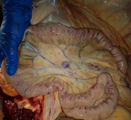
Gross findings: Findings were inconclusive as autolysis had already started, although swelling and some dark areas in the muscles raised the possibility of clostridial diseases. A fresh post mortem was needed to confirm (or rule out) the cause. This was done four days after the initial inquiry on a very sick bull from the third consignment. Gross pathology included port wine coloured urine, an enlarged gall bladder and petechial haemorrhages on numerous mucosal surfaces (see photos).
The differential list included: tick fever (a 2-week incubation with losses after trucking being consistent with naïve animals moving into a tick zone); clostridial disease; botulism from pre-formed toxin in feed; and a poisoning like ironwood. Tick fever was looking likely although spleen involvement and jaundice was not as evident as expected.
Laboratory findings: A low red cell count, haemoglobin and platelets were found on haematology. A mild regenerative anaemia and occasional erythrocytes containing intracellular organisms consistent with Babesia bovis were seen on the blood smear. The tick fever diagnosis was confirmed by a brain smear (see image which shows red blood cells in a capillary, with many containing pear-shaped, often paired, basophilic organisms consistent in size and shape with Babesia bovis).
Recommendations to the producer: Normally cattle being transferred to more northern properties are vaccinated with 3 germ (allowing sufficient time for protection to develop).Options in this case:
- Send cattle direct from original property to the holding yard.
- Put cattle in yards on the intermediate property on hay if holding for few days.
- Treat with Imidol to give 4 weeks cover (too expensive for large number of commercial cattle).
- Dip with a chemical that has residual effect (eg Bayticol to keep ticks off for up to 10-12 days).
Reflections of the vet:
- The time interval set alarm bells ringing – I had seen this happen years before with animals from a clean property via a property in tick zone .
- The challenge was not to jump exclusively onto tick fever without considering other differentials.
- The lack of evidence from the early post mortems was a little off-putting as this usually provides good evidence of tick fever – but the fact that some autolysis had set in probably explains some of this.