Septicaemia with multi-organ vasculitis and thrombosis Unusual presentation: Sixteen deaths in a mob of young bulls within a month of arriving on a new property. Above: A depressed, non-responsive bull – with ticks but no signs of ocular or nasal discharge....
Gross path challenge #5 Photo: Ayrial Foster, Berrimah DPIR NT BOVINE LUNG: The visceral pleural surface displays moderate, regionally extensive opacity most obvious over the dorsocaudal region. Diffusely, the interlobular pulmonary septae are obvious and expanded by...
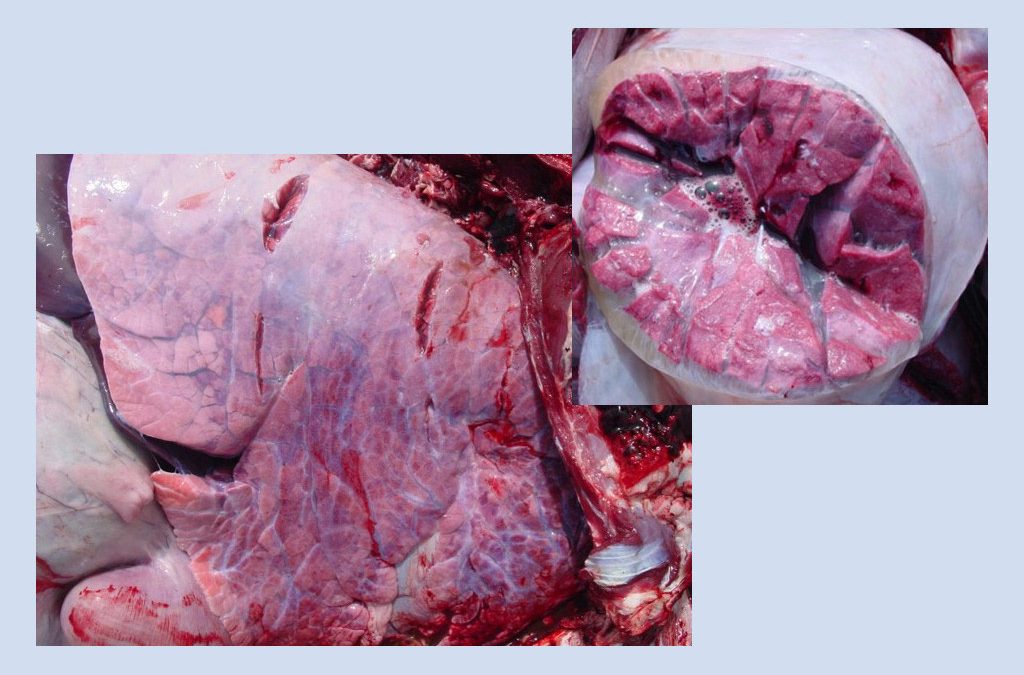
Gross path challenge #3 Photo: The Joint Pathology Centre WSC 2015 SKELETAL MUSCLE: The cut surface of the muscle shows large multifocal to coalescing dark red to black areas that are dry. Occasional gas bubbles (emphysema) are evident in the interstitial connective...
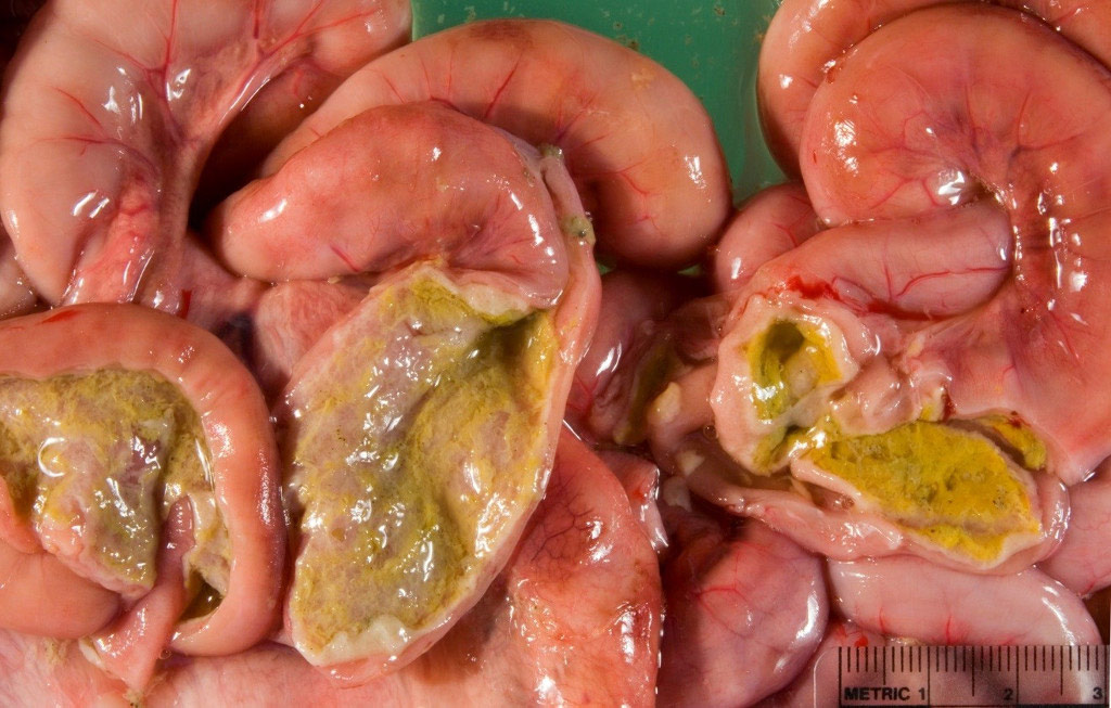
Gross path challenge #2 Photo: The Joint Pathology Centre WSC 2009 SMALL INTESTINE: The wall of the small intestine is thickened and the serosal surface is diffusely dark pink. The mucosa is coated with yellow friable material. Dx: Enteritis, salmonellosis Back to...
Gross path challenge #1 Photo: The Joint Pathology Centre WSC 2012 LIVER: The cut surface of the liver is mottled with depressed irregular bright red areas (1-2mm diameter) surrounded by light brown areas (accentuated lobular pattern). Dx: Acute hepatic necrosis,...